Nanoscale Imaging
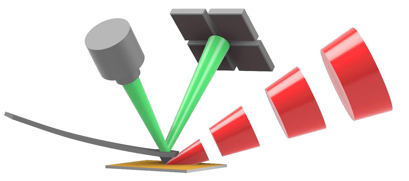
In our nearfield lab we take advantage of photothermal infrared (IR) spectroscopy. Chemical imaging at nanoscale spatial resolution is enabled with scanning probe microscope IR spectroscopy (AFM-IR).

Motivation
Traditional far-field IR microscopes are limited in spatial resolution by the diffraction limit, which prevents the achievement of resolutions below several micrometres. This prevents the analysis of objects below this size, such as most intracellular structures, interfaces, crystals, and proteins, to name a few. To circumvent this problem, a technique called AFM-IR that combines atomic force microscopy (AFM) with infrared spectroscopy (IR) was developed. In this technique, a pulsed IR laser source aimed at the area bellow the AFM cantilever provides IR radiation which can be absorbed by the sample. The resulting thermal expansion is detected by the highly sensitive AFM cantilever. Thus, the detection occurs in the near-field and circumvents the diffraction limit. AFM-IR can reach resolutions of down to 20 nm in contact mode and 10 nm in tapping mode.
Areas of Research
New Applications
In our work, we apply AFM-IR to a broad range of samples/areas of research:
1In the last few years, we have developed an approach to intracellular protein detection in fungi through the combination of AFM-IR, fluorescence microscopy and chemometric models. AFM-IR is able to detect cell components without the need for labeling or staining, purely from spectra and chemometrics.
2When applied to polymeric materials AFM-IR reveals main components, phase distribution and contaminants. We have established a protocol for tapping mode AFM-IR of polymers. And recently we used AFM-IR to study a recycled post-consumer polyolefin blends. The data that AFM-IR provides helps polymer scientists to develop better materials and achieve higher recycling rates.
3Other, recent areas of interest include energy materials and plant tissues.
New Methods for AFM-IR
Currently, we are working on developing instrumentation to allow for simpler measurements in liquid, as well as gaining a deeper understanding of the characteristics of the technique through the finding of a point spread function.
Researchers
Georg Ramer
Margaux Petay
Elisabeth Holub
Lena Neubauer
Nikolaus Hondl
Sebastian Wöhrer
Yide Zhang
Publications
- U. Yilmaz, S. Sam, B. Lendl, G. Ramer, “Bottom-Illuminated Photothermal Nanoscale Chemical Imaging with a Flat Silicon ATR in Air and Liquid” Anal. Chem. 2024, doi: doi.org/10.1021/acs.analchem.3c04348.
- M. Wang et al., “High Throughput Nanoimaging of Thermal Conductivity and Interfacial Thermal Conductance,” Nano Lett., vol. 22, no. 11, pp. 4325–4332, Jun. 2022, doi: 10.1021/acs.nanolett.2c00337.
- A. C. V. D. dos Santos, B. Lendl, and G. Ramer, “Systematic analysis and nanoscale chemical imaging of polymers using photothermal-induced resonance (AFM-IR) infrared spectroscopy,” Polymer Testing, vol. 106, p. 107443, Feb. 2022, doi: 10.1016/j.polymertesting.2021.107443.
- M. Weiss et al., “Elemental mapping of fluorine by means of molecular laser induced breakdown spectroscopy,” Analytica Chimica Acta, vol. 1195, p. 339422, Feb. 2022, doi: 10.1016/j.aca.2021.339422.
- A. C. V. D. dos Santos, D. Tranchida, B. Lendl, and G. Ramer, “Nanoscale chemical characterization of a post-consumer recycled polyolefin blend using tapping mode AFM-IR,” Analyst, vol. 147, no. 16, pp. 3741–3747, 2022, doi: 10.1039/D2AN00823H.
- M. Winzely et al., “AFM investigation of APAC (antiplatelet and anticoagulant heparin proteoglycan),” Anal Bioanal Chem, Nov. 2021, doi: 10.1007/s00216-021-03765-y.
- A. C. V. D. dos Santos et al., “Nanoscale Infrared Spectroscopy and Chemometrics Enable Detection of Intracellular Protein Distribution,” Anal. Chem., Dec. 2020, doi: 10.1021/acs.analchem.0c02228.
- G. Ramer et al., “High-Q dark hyperbolic phonon-polaritons in hexagonal boron nitride nanostructures,” Nanophotonics, vol. 9, no. 6, May 2020, doi: 10.1515/nanoph-2020-0048.
- X. Ma et al., “Revealing the Distribution of Metal Carboxylates in Oil Paint from the Micro‐ to Nanoscale,” Angew. Chem., vol. 131, no. 34, pp. 11778–11782, Aug. 2019, doi: 10.1002/ange.201903553.